J.H.JIN의 모든 글

Thermo plate

TOKAI HIT Thermo Plate Clear Glass Heater

현미경 관찰하는 동안 시료의 정확하고 안정적인 열 제어 보장.

생명공학분야에서 산업분야에 이르기까지 다양한 종류의 온도 제어.

TPi 시리즈는 이전 버전에 비해 탁월한 성능과 새로운 기능으로 설계된 제품입니다

Glass Heater(TPi-110RX) & Controller
  • Stereo, Uplight, Inverted Microscope에 장착하여 정확한 온도조절 가능
  • 강화유리 장착으로 다양한 실험에서도 무리 없이 사용 가능
  • 강화유리 10년 무상 보증
  • Plate에 Green LED장착으로 최적의 온도 상태 표시.
  • 유리 전체에서 균일하게 가열할 수 있는 특허기술로 샘플전체에 일정한  온도 조절 가능
  • Nikon, Olympus, Leica, ZEISS에 맞는 제품 공급 가능

Standard Type(Glass Heater) 제품의 특징

  • 강화유리 적용으로 파손에 대한 걱정이 없습니다

TPi 시리즈 Thermoplate의 70% 이상이 강화유리 히터를 사용하며 Glass 파손에 대한 10년 무상 수리 서비스를 제공 합니다.

참고: 모델명이 “X”로 끝나는 모델이 강화 유리 적용 모델 입니다. (TPi-UNIX → 강화유리 적용 모델)

  • LED 표시장치
LED표시로 Plate 상태를 쉽게 알수 있습니다.
  • Compact한 디자인과 손쉬운 설치
W85 x D135 x H30 mm, 약 2Kg.
마운팅 후크 장착으로 controller 각도를 조절하여 테이블 또는 벽에 걸어서 사용 가능

작고 가벼운 설계로 되었으며, Muli-Functional 시스템은 다양한 분야에서 정확한 온도 조절을 제공 합니다.

  • 쉬운 온도 측정
K-type Thermocouple

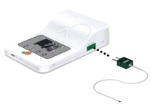
멸균센서를 사용하여 샘플의 실제온도 또는 플레이트 표면의 온도를 측정 할 수 있습니다.

  • 온도 관리 프로그램 TEM
CVS data 저장/ 온도 보정 기능 포함

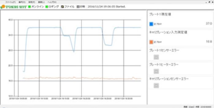
플레이트 표면의 온도, 설정값 및 외부 온도 센서를 모니터링.

CVS 형식으로 저장가능.

Thermo Plate TPi 시리즈 기본 제공.

  • 원터치 온도 보정
Calibration button

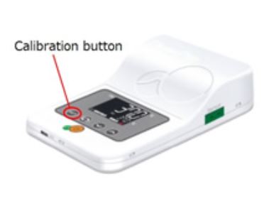
실내 온도에서 시스템의 온도를 재 보정 할 수 있습니다.

출하시 실내온도 37℃ 에서 출하시 25 ℃ ± 2 ℃ 로 설정 되어있습니다.

  • 균일한 온도 전달

Thermo Plate / Thermo Stage / Warming Plate / Heating Plate / Heating Stage / 가열 스테이지


아래 링크에서 현미경 제조사별 모델 검색이 가능합니다.

Touptek camera

Entry Level MicroScope USB2.0 Camera UCMOS Series ToupTek Photonics Co., Ltd

USB2.0 인터페이스 CMOS 카메라

USB2.0 인터페이스 CMOS 카메라로 저렴한 비용으로 탁월한 현미경영상 획득에 용이합니다.

실체현미경, 생물 현미경, 금속 현미경등 대부분의 현미경에 적용 가능합니다.

  • Aptina CMOS 센서 장착.
  • 0.35M ~ 14M 까지 다양한 해상도 선택 가능.
  • USB2.0 인터페이스로 고속 데이터 전송.
  • Ultra-Fine color engine 장착으로 완벽한 색 재현 가능.
  • 정지 이미지, 동영상 촬영 가능.
  • IR cut 필터 내장
  • PC USB 전원공급.
  • Windows XP / Vista / 7 / 8 / 10 (32&64bit) , OSx, Linux 사용가능.

High Level MicroScope USB3.0 CMOS Camera E3CMOS / E3ISPM Series

USB3.0 High Performance CMOS Camera E3CMOS Series

USB3.0 인터페이스 CMOS 카메라로 저렴한 비용으로 탁월한 현미경영상 획득에 용이합니다.

형광현미경을 포함한 생물 현미경, 고성능 금속 현미경등 대부분의 현미경에 적용 가능합니다.

  • Sony Exmor CMOS 센서 장착.
  • 0.4M ~ 20M 까지 다양한 해상도 선택 가능.
  • USB3.0 인터페이스로 5Gbps 고속 데이터 전송.
  • Ultra-Fine color engine 장착으로 완벽한 색 재현 가능.
  • 정지 이미지, 동영상 촬영 가능.
  • IR cut 필터 내장
  • PC USB 전원공급.
  • Windows XP / Vista / 7 / 8 / 10 (32&64bit) , OSx, Linux 사용가능.

HDMI출력 , 고성능 CMOS 칩을 장착한 Touptek XCAM1080 series입니다.

Touptek XCAM1080 시리즈

Touptek XCAM1080 시리즈는 HDMI, USB2.0, SD card , WiFi(PHB모델) 다양한 출력 방식으로 적용이 가능합니다.

  • 적용분야
  • 기초과학 연구
  • 교육 (강의, 학술교류)
  • 디지털 실험실
  • PCB, 회로 검사실
  • 병리학 실험실
  • 미생물관찰
  • 우주항공, 군사용

HDMI 방식을 이용할 경우 PC 없이 모니터에 직접 연결하여 실습교육, 현장 외관검사용으로 , 고해상도의 이미지 획득/검사/저장이 가능합니다

IRIS 9 HIGH RESOLUTION IMAGING

Large Field of View sCMOS Camera

High Resolution Imaging IRIS 9

Iris 9 Scientific sCMOS camera는 Live cell Imaging에 최적화된 고해상도 카메라입니다.

  • Iris 9 Scientific camera
  • 75% Quantum Efficiency
  • 4.25um x 4.25um Pixe Area
  • 9 Megapixel (2960×2960)

Applications

Customer Stories

Specification

Sensor TechnologysCMOS, Gpixel GSense 5130 Scientific CMOS sensor
Quantum Efficiency73%
Pixel Size4.25um X 4.25um
Supported InterfacesPCIe ,USB3.0
Spectrum CapabilitiesVisible(400-700nm), Near Infrared(700-1000nm)
Read Noise(median)1.5 e-
Sensor Area12.61mm X 12.61mm , 17.8mm Diagonal
Frame Rate30fps @16bit
Iris 99M pixels (2960×2960)
Lens InterfaceC-mount

Resolution and Pixel Size

The small, 4.25 µm pixels provide highly detailed images across the imaging plane, which allows for the highest resolution when using lower magnification objectives.

sCMOS Sensitivity

The low noise and high quantum efficiency across a wide range of wavelengths produces higher quality images with lower exposure times than conventional CCD cameras.

Advanced Triggering

Programmable Scan Mode provides increased control over the rolling shutter exposure and read-out functionality of CMOS sensors by providing access to the sensor timing settings to allow optimization around applications that require control over the line time.

Superior Background Quality

The Iris 9 features Pattern Noise Reduction Technology and Correlated Noise Reduction Technology to ensure that it delivers clean, pattern-free images with minimal pixel defects, delivering improved image quality in low light conditions.

Compact Form Factor

The Iris 9 is a compact 76 x 76 x 88 mm with optimized cooling for the size, ideal for integration into new or existing configurations.

IRIS 9 Camera Datasheet

Iris Camera Manual

DIC imaging of the cardinal vein of a zebrafish taken on the Iris 9 scientific CMOS.
Zebrafish cilia captured at 100 fps on the Iris 9 Scientific CMOS camera

IRIS 15 High Resolution Imaging

High Resolution Imaging IRIS15

IRIS15 Scientific sCMOS 카메라는 넓은 FOV와 15M pixels의 고해상도 이미지를 제공 합니다.

  • IRIS15 Scientific camera
  • 15Megapixel Camera (5056×2960)
  • 4.25um x 4.25um Pixel Area
  • Large 25mm Diagonal Field of View

Applications

Customer Stories

Specification

Sensor TechnologysCMOS, Gpixel GSense 5130 Scientific CMOS sensor
Quantum Efficiency73%
Pixel Size4.25um X 4.25um
Supported InterfacesPCIe ,USB3.0
Spectrum CapabilitiesVisible(400-700nm), Near Infrared(700-1000nm)
Read Noise(median)1.5 e-
Sensor Area21.49mm X 12.61mm , 24.9mm Diagonal
Frame Rate30fps @16bit
Iris 1514M pixels (5056×5056)
Lens InterfaceF-mount

Large Field of View

The larger format 25 mm sensor of the Iris 15 is designed to increase throughput, maximize the amount of data captured and take full advantage of new, larger field of view microscopes.

High Resolution

The small, 4.25 µm pixels provide highly detailed images across the imaging plane, which allows for the highest resolution when using lower magnification objectives.

Advanced Triggering

Programmable Scan Mode provides increased control over the rolling shutter exposure and read-out functionality of CMOS sensors by providing access to the sensor timing settings to allow optimization around applications that require control over the line time.

Superior Background Quality

The Iris 15 features Pattern Noise Reduction Technology and Correlated Noise Reduction Technology to ensure that it delivers clean, pattern-free images with minimal pixel defects, delivering improved image quality in low light conditions.

Compact Form Factor

The Iris 15 is a compact 78 x 78 x 108 mm with optimized cooling for the size, ideal for integration into new or existing configurations.

IRIS 15 Camera Datasheet

Iris Camera Manual

Kinetix scmos scientific camera

Kinetix High Speed, Back-illumnated

Back-illuminated Kinetix Scientific sCMOS 카메라는 가장 균형잡힌 Pixel 크기와 완벽한 95%QE효율로 가장 빠른 속도와 최대 FOV를 제공 합니다.

  • 400 FPS
  • 29.4mm Diagonal FOV
  • 1.2 e- Read Noise
  • 95% Quantum Efficiency
  • 6.5um x 6.5um Pixel Area
  • 3200×3200 (10.24Megapixel)

Applications

Customer Stories

Specification

Sensor TechnologysCMOS
Quantum Efficiency95%
Pixel Size6.5um X 6.5um
Supported InterfacesPCIe and USB3.2
Spectrum CapabilitiesUV(200-400nm), Visible(400-700nm), Near Infrared(700-1000nm)
Read Noise(median)1.2 e-
Sensor Area20.8mm X 20.8mm , 29.4mm Diagonal
Frame Rate400fps @ 8bit / 90fps @12bit / 83fps @ 16bit
Kinetix10.2M pixels (3200×3200)

Extreme Speed

Taking advantage of an 8-bit readout mode, the Kinetix sCMOS delivers a tremendous 400 frames per second (fps), full frame with a 29.4 mm diagonal field of view. The optimized line time allows the speed to significantly outperform typical sCMOS devices, delivering over 4000 megapixels/second – an almost 10-fold improvement

Large Field Of View

The 29.4 mm square sensor of the Kinetix is designed to increase throughput, maximize the amount of data captured in a single frame and take full advantage of new, larger field of view microscopes. At 29.4 mm diagonal, the Kinetix sensor has a 2.4x larger imaging area than typical sCMOS cameras allowing the user to significantly speed up data acquisition.

High Sensitivity

The Kinetix back-illuminated sCMOS camera achieves a near-perfect 95% quantum efficiency. By bringing the light in from the back of the sensor, photons land directly onto the light receiving surface, maximizing light collecting capability. The Kinetix combines 95% quantum efficiency with a low 1.2 e- read noise to deliver the most sensitive sCMOS camera at over 400 frames per second.

High Resolution

The Kinetix features 6.5 µm x 6.5 µm pixels, the accepted standard for most live cell applications using 40x and 60x magnification. This pixel size provides highly detailed images across the imaging plane and is most suitable for the broadest range of microscope objectives.

Superior Background Quality

The Kinetix features Pattern Noise Reduction Technology and Correlated Noise Reduction Technology to ensure that it delivers clean, pattern-free images with minimal pixel defects, delivering improved image quality in low light conditions.

Kinetix sCMOS Camera Datasheet (Preliminary)

PRIME BSI express SCIENTIFIC SCMOS CAMERA

Prime BSI EXPRESS

Prime BSI Express Scientific CMOS(sCMOS)는 최적화된 컴팩트 플랫폼으로 최적화된 Pixel 디자인, USB 3.2 Gen 2 연결성, 완벽한 95% Quantum Efficiency를 통해 고해상도 고감도 이미징의 완벽한 균형을 제공함으로써 최대한의 신호를 감지합니다.

  • Prime BSI EXPRESS
  • 95% Quantum Efficiency
  • 1.0e- Read Noise
  • 6.5um x 6.5um Pixel Area
  • 95 FPS over USB3.2 Gen2

Applications

Customer Stories

Specification

Sensor TechnologysCMOS
Quantum Efficiency95%
Pixel Size6.5um X 6.5um
Supported InterfacesUSB3.2 Gen2
Spectrum CapabilitiesUV(200-400nm), Visible(400-700nm), Near Infrared(700-1000nm)
Read Noise(median)1.0 e-
Sensor Area13.3mm X 13.3mm
Frame Rate43fps @ 16bit / 43fps @12bit / 95fps @11bit
Prime BSI Express4.2M pixels (2048×2048)

Sensitivity

By bringing the light in from the back of the sensor, photons land directly onto the light receiving surface, maximizing light collecting capability. The Prime BSI Express combines 95% quantum efficiency with the low 1.0 e- read noise CMS mode to deliver the most sensitive camera based on sCMOS technology at over 40 frames per second.

Compact Form Factor

The Prime BSI Express is a compact 78mm x 78mm x 90mm with optimized cooling for the size, ideal for integration into new or existing configurations.

Interface and Integrate

Interfacing and integration is simple with the Prime BSI Express, which uses USB 3.2 Gen 2 for truly uncomplicated connectivity. Our standard PVCAM driver can be easily installed in Windows operating systems to get the Prime BSI Express running in minutes.

Resolution and Pixel Size

The Prime BSI Express features 6.5 µm pixels, the accepted standard for most live cell applications. This pixel size provides highly detailed images across the imaging plane and is most suitable for the broadest range of magnifications.

Superior Background Quality

The Prime BSI Express features Pattern Noise Reduction Technology and Correlated Noise Reduction Technology to ensure that it delivers clean, pattern-free images with minimal pixel defects, delivering improved image quality in low light conditions.

Prime BSI Express Camera Datasheet

Prime BSI Express Camera Manual

RETIGA ELECTRO : DESIGNED FOR ELECTROPHYSIOLOGY

Retiga Electro : Designed for Electrophysiology

Retiga Electro : Designed for Electrophysiology

​● CCD Array: 1360 x 1024 pixels (6.45um x 6.45um)

​● Sensor Dimension : 8.8 mm x 6.6mm (11mm)

​​● Frame Rate: 22 fps (Full resolution), 31 fps (binning 2×2)

● 1.4MP 75% QE 효율 CCD Camera로 Extreme Low Light Imaging에 적합

● USB 3.0:50MHz high frame rate를 적용하여 이미지의 끊김 없이 포커스 조절이 가능하며, 빠르게 원하는 샘플을 찾을 수 있음

● DPC(Defective Pixel Correction)을 적용하여 열화 된 픽셀을 보정하고, 선명한 영상 구현

<Application>

Advanced Technical Features

The advanced technical features of the Retiga ELECTRO were designed to empower electrophysiologists to image without exogenous noise that pollute recordings. This is accomplished by coupling regulated fanless cooling and external grounding with FPGA-based intelligent features to correct defective pixels.

High Sensitivity

The Retiga ELECTRO features 75% peak quantum efficiency (QE) and maintains a high QE across commonly used wavelengths in electrophysiology imaging. This is combined with low noise electronics to reveal weakest fluorescence signals. The Retiga ELECTRO also has an exceptionally low dark current so long stare applications will not suffer in
image quality.

Simple Installation

The Retiga ELECTRO delivers data over the simple, versatile and high-speed USB 3.0 interface. This standard, accepted interface is present on most modern computers, simplifying connectivity. The Retiga ELECTRO is also supported on the latest Windows operating systems ensuring modern system compatibility.

Retiga ELECTRO Camera Datasheet

Retiga ELECTRO Camera Manual

EVOLVE 512 DELTA EMCCD CAMERA

Evolve 512 Delta EMCCD Camera

New LightSpeedTM Mode

  – 최대 2400 FPS실현

  – Dual Channel Imaging기술을 적용 두파장의 형광을 한 카메라에서 고속으로 획득 가능.  

● 20MHz 512×512 EMCCD

● 67 Frames per Second.

● 72e- Read Noise, <1e- Using EM Gain

● > 90% Quantum Efficiency

● Quantitative Performance

● Includes full two year warranty

<Application>

■ Super-Resolution Microscope

■ FCS, TIRF, FRET, FRAP

■ Spining Disk Confocal

■ SPIM

■ Intrinsic Image

Evolve 512 Delta DataSheet

Evolve 512 Delta Manual

QIMAGING RETIGA R6 CCD CAMERA

Qimaging Retiga R6 CCD Camera

​● CCD Array: 2688 x 2200 pixels (4.54um x 4.54um)

​● Sensor Dimension : 12.5 mmx 10mm (16mm)

​​● Frame Rate: 7.1 fps (Full resolution), 12.8 fps (binning 2×2)

● 75% QE 효율 CCD Camera로 Extreme Low Light Imaging에 적합

● USB 3.0:50MHz high frame rate를 적용하여 이미지의 끊김 없이 포커스 조절이 가능하며, 빠르게 원하는 샘플을 찾을 수 있음

● Real-time FPGA의 알고리즘을 적용하여 깨끗한 영상 구현

<Application>

■ Live cell Imaging and Fixed cell detecton

​■ Time Lapse Imaging 

■ Tile-and Stitch Microscopy

■ Stereo Microscopy

Large Field Of View

The Retiga R6 delivers a large, 16 mm diagonal field of view designed to increase throughput and maximize the amount of data captured in a single frame. Image larger samples or increase throughput by imaging multiple cells at once by taking advantage of a larger field of view.

High Resolution

The Retiga R6 features 6 million, 4.54 µm pixels to maximize the resolving power of your system. The small pixels provide highly detailed images across the imaging plane, which allows for the highest resolution when using lower magnification objectives.

Simple Installation

The Retiga R6 delivers data over the simple, versatile and high-speed USB 3.0 interface. This standard, accepted interface is present on most modern computers, simplifying connectivity. The Retiga R6 is also supported on the latest Windows operating systems ensuring modern system compatibility.

Retiga R6 Camera Datasheet

Retiga R6 Camera Manual

PRIME BSI SCIENTIFIC sCMOS CAMERA

Prime BSI

Prime BSI Scientific sCMOS 카메라는 최적화된 Pixel 디자인과 95 % Quantum Efficiency로 고해상도, 고감도 이미징의 완벽한 균형을 제공합니다.

  • 95% Quantum Efficiency
  • 6.5um x 6.5um Pixel Area
  • 63 FPS

Specification

Sensor TechnologysCMOS
Quantum Efficiency95%
Pixel Size6.5um X 6.5um
Supported InterfacesPCIe and USB3.0
Spectrum CapabilitiesUV(200-400nm), Visible(400-700nm), Near Infrared(700-1000nm)
Read Noise(median)1.0 e-
Sensor Area13.3mm X 13.3mm
Frame Rate43fps @ 16bit / 63fps @12bit
Prime BSI4.2M pixels (2048×2048)

<Application>

Customer Stories

Sensitivity

The Prime BSI back-illuminated sCMOS camera achieves a near-perfect 95% quantum efficiency. By bringing the light in from the back of the sensor, photons land directly onto the light receiving surface, maximizing light collecting capability. The Prime BSI combines 95% quantum efficiency with a low 1.0 e- read noise to deliver the most sensitive camera based on sCMOS technology at over 40 frames per second.

Resolution and Pixel Size

The Prime BSI features 6.5 μm pixels, the accepted standard for most live cell applications using 40x and 60x magnification. This pixel size provides highly detailed images across the imaging plane and is most suitable for the broadest range of microscope objectives.

Superior Background Quality

The Prime BSI features Pattern Noise Reduction Technology and Correlated Noise Reduction Technology to ensure that it delivers clean, pattern free images with minimal pixel defects, delivering improved image quality in low light conditions.

Advanced Triggering

Programmable Scan Mode provides increased control over the rolling shutter exposure and read-out functionality of CMOS sensors for applications that require control over the line time. Sequenced Multiple Acquisition Real Time Streaming (SMART) Streaming enables the Prime BSI to capture a continuous sequence of images while cycling through 16 pre-programmed exposure time values, delivering the fastest multi-channel acquisition times.

Advanced Features

The Prime BSI offers PrimeEnhance active denoising which provides a real-time quantitative increase in the signal to noise ratio to maximize sensitivity at ultra-low light levels.

Prime BSI Camera Datasheet

Prime BSI Camera Manual

BSI Handbook