CoolLED pE-300ultra
NOW DOUBLE THE INTENSITY AT THE SAMPLE PLANE
CoolLED’s pE-300ultra is a fluorescence microscopy light source which offers intense, broad-spectrum LED illumination for imaging most common fluorescent stains. Users have access to both microsecond switching via multiple TTL inputs and the ability to mount inline excitation filters. This, when paired with today’s high performance multi band filter sets, facilitates imaging traditionally done via a white light source and a filter wheel, with all the benefits of LED.


The pE-300ultra is the most controllable member of the pE-300 Series. In addition to the many features of the pE-300lite and the pE-300white, the system offers precise control over wavelength intensity and shuttering. Until now these benefits have only been accessible to users of high end, highly flexible illuminators such as the CoolLED pE-4000.
Throughout 2017 we will be releasing exciting new products and product updates. The first member of the CoolLED product range to receive the star treatment is the pE-300 Series.
As part of the award winning pE-300 Series, the NEW Enhanced pE-300ultra now delivers DOUBLE the intensity at the microscope sample plane. It allows adjustment of output in 1% steps, giving precise control. The broad spectrum covers everyday fluorophores: DAPI, CFP, Aqua, FITC, TRITC, TxRed, Cy5 and many more.

It is now possible to view and image your samples brighter than ever.

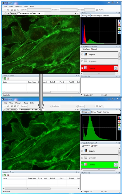
Convaleria taken with Photometrics Prime camera with 10ms exposure.
LED illumination means no mercury to dispose of and lower energy costs due to low power consumption and the precise control offered. With the new enhanced pE-300ultra, all 3 channels at full power use just 46 Watts. That is almost a quadruple efficiency increase!
This compares to other LED technology that uses 120 to 350 Watts. This leap forward in technology makes LEDs more attractive when compared to old Mercury or Metal Halide technology. Where “green” funding is available, the reduction in institutional energy usage improves the return on investment.
CoolLED’s pE-300 Series was the 2017 winner of the “Go Beyond Award” in the “Products” category for “Excellence in sustainability in laboratory and other high technology facilities” The International Institute for Sustainable Laboratories (I2SL) runs a unique awards program honouring organisations, individuals, products,and projects that are advancing sustainable, high-performance facilities.
The pE-300ultra offers triggering from multiple TTL inputs which, coupled with the ability to mount inline excitation filters, provides microsecond switching of pre-filtered excitation light. This, when paired with today’s high performance multi-band filter sets, facilitates imaging traditionally done using a white light source and a filter wheel, with all the benefits of LED, and most excitingly at speeds not previously so affordable.

The system comprises a pE-300ultra Light Source, Control Pod, set of three Excitation Filter Holders and Power Supply. It can be specified with a microscope adaptor for direct fit configuration or with a 3mm diameter liquid light guide if there is a need to keep the source of illumination remote from the microscope. An optional pE-Universal Collimator and microscope adaptor can be selected for use with a liquid light guide.
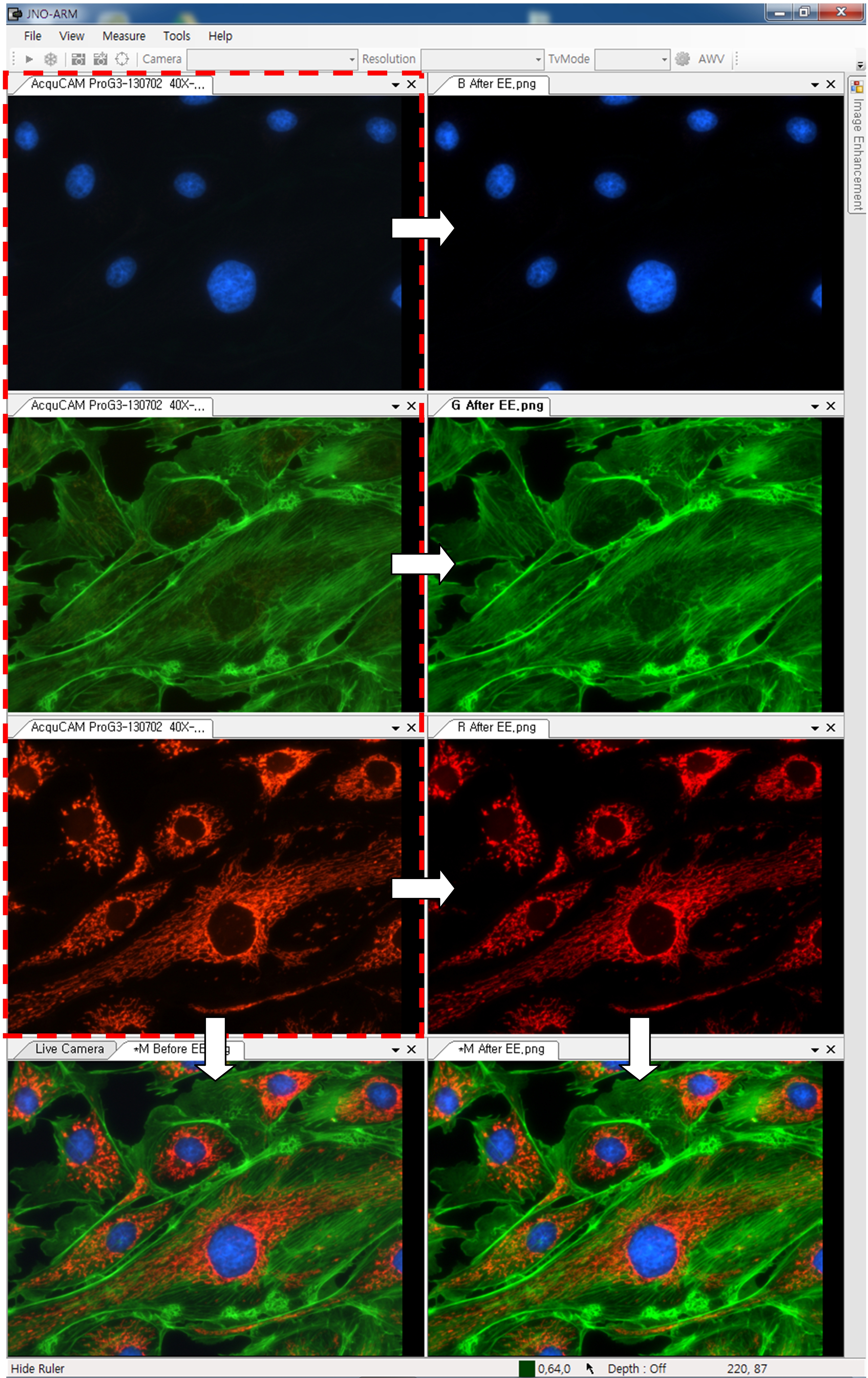
A benefit of the pE-300ultra is that stains can be viewed either individually or in combination, without filter cube changes. This makes it ideal for use with multi-band filter sets as the screening process can be simplified when fewer filter cubes are used. Independent control of the three LED channels means that the user can control the level of excitation of each fluorescent stain independently on a multi-stained sample, potentially removing the need for single band filter sets altogether. For further information on improved multi-stain testing, go to the Downloads tab.
An important additional feature of the pE-300ultra is that it includes CoolLED’s “Sequence Runner” multiple channel excitation mode. Users can define the order of their fluorophore capture using their pE-300ultra Control Pod, then the pE-300ultra light source can accept a single TTL output from the experiment set-up’s camera to initiate the step-through of a sequence of excitation channels. This feature is independent of the individual channel TTL inputs on the light source. This offers users the facility to run through a sequence of excitation channels using a camera which has only a single TTL-out.
With an expected lifetime in excess of 25,000 on hours and a comprehensive range of microscope adaptors, the pE-300ultra can be fitted to most current and older microscopes and operate for many years without aligning or replacing bulbs. The result is a safe, convenient illumination system without any additional operating costs.

There are two pE-300ultra configuration options:
Direct-fit for connecting to a microscopes – by selecting from a range of microscope adaptors which covers all current and most older models. A simple once only adjustment will allow optimisation to the optical path of the microscope.
Liquid Light Guidewith a fixed 3mm diameter, liquid light guide. An optional pE-Universal Collimator can be specified in conjunction with a microscope adaptor if required.
The pE-300ultra offers:
- Clean bright illumination across the spectrum – excites common fluorescent stains
- Specify for existing single and multi-band filter sets – no need to buy new filters
- Instant on/off – No shutters required, no warm up or cool down
- Simple to fit, simple to use – no alignment, a once only adjustment
- Stable & repeatable – reliable and consistent results
- Wide range of microscope adaptors – fits most microscopes
- Precise intensity control in 1% steps (0-100%) – no ND filters required
- Excellent uniformity over field of view – fixed and stable, no alignment necessary
- Long lifetime – expected to exceed 25,000 hours of operating time
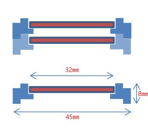
- Removable inline excitation filter holders – no moving parts
- Individual channel triggering via TTL in microseconds
- Sequence Runner – sequenced excitation from a single TTL-out